14
9985-1000311
9985-1000247
9985-1005532
9985-1000260
9985-1000312
Anatomy
3 B S c i e n t i f i c ® M e d i c a l
Hi s tology
3B MICRO
anatomy
™ Eye
This model illustrates the microscopic structure of the retina with choroid
and sclera. The left block like, layered side of the model side shows the
complete structure of the retina including the vascular layer and parts of
the sclera from a light microscopic view. The right part of the model is a
sectional enlargement. It shows the microscopic structure of the photore-
ceptors and the cells of the pigmented layer.
25x23x18.5 cm; 1.2 kg
&
L/D/E/F/S/P/I/J www.
9985-1000260
3B MICRO
anatomy
™ Tongue
The latest model in our 3B MICRO
anatomy
™ series, the tongue, is fascinat-
ing because it combines enlargements of various different parts of the
tongue in one model. It comprises a macroscopic view of the tongue in life
size (dorsal view) and microscopic views of the various papillae of the
tongue (10-20x life size) and of a taste bud (approx. 450x life size). All views
are mounted on a base that also features an overview of the sensory and
sensitive innervation of the tongue. A unique model for an intensive study
of the tongue.
14.5x32.5x20 cm, 0.8 kg
&
L/D/E/F/I/S/P/J/R/C www.
9985-1000247
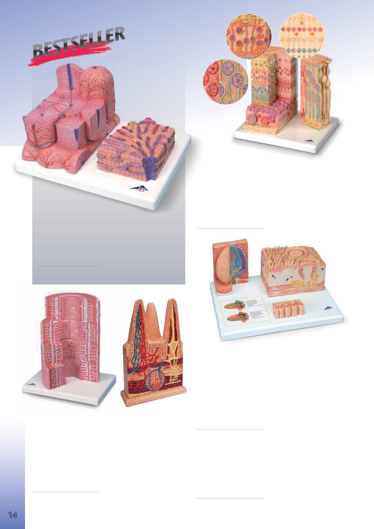
3B MICRO
anatomy
™ Digestive System
The model illustrates the structure of the fine tissues of four characteristic
sections of the digestive system: oesophagus, stomach, small intestine,
large intestine. The front of the model, from top to bottom, shows a magni-
fied view in histological section of the individual sections of the digestive
system and their fine tissue structures.On the back of the model, highly
magnified views of didactically interesting areas of each of the digestive
system sections shown on the front are emphasized.
29.5x26x18.5 cm; 1.5 kg
&
L/E/D/S/F/P/I/J www.
9985-1000311
3B MICRO
anatomy
™ Liver
This 2-part model shows a highly magnified diagrammatic view of a
section of the liver. The left part of the model shows a section of the
liver that comprises several lobules. The right part of the model is a
highly magnified view of the sectioned lobule on the left.
15x26x18.5 cm; 0.7 kg
&
L/E/D/S/F/P/I/J www.
9985-1000312
Intestinal Villi, 100 times life-size
This model consists of one entire villus, one longitudinally sectioned villus
showing the arterioles and venules and one sectioned villus to show the
lymphatic vessels. Also includes a longitudinal section of Lieberkühn’s
crypt. On base.
43x28x10 cm; 2.5 kg
9985-1005532


