15
9985-1000290
9985-1000213
9985-1000289
9985-1000301
9985-1000154
9985-1000291
. . . g o i n g o n e s t e p f u r t h e r
Anatomy
Hi s tology
3B MICRO
anatomy
™ Kidney
This extremely detailed model shows the morphologic / functional units of
the kidney greatly magnified. Six model zones illustrate the following fine-
tissue structures that serve the production of urine:
• Longitudinal section of a kidney
• Section of renal cortex and renal medulla
• Wedge-shaped section of a kidney lobe with a diagrammatic depiction of
three nephrons with Henle’s loops of different lengths and diagrammatic
depiction of the vascular supply
• Diagrammatic illustration of a nephron with a short Henle’s loop and
didactic / diagrammatic illustration of the vascular supply
• Diagrammatic illustration of an opened renal corpuscle with nephron
and light-microscopic transverse sections of the proximal, attenuated and
distal segments of a renal tubule
• Diagrammatic / didactic illustration of an opened renal corpuscle
Mounted on a base.
23.5x25.5x19 cm; 1.3 kg
&
L/E/D/S/F/P/I/J www.
9985-1000301
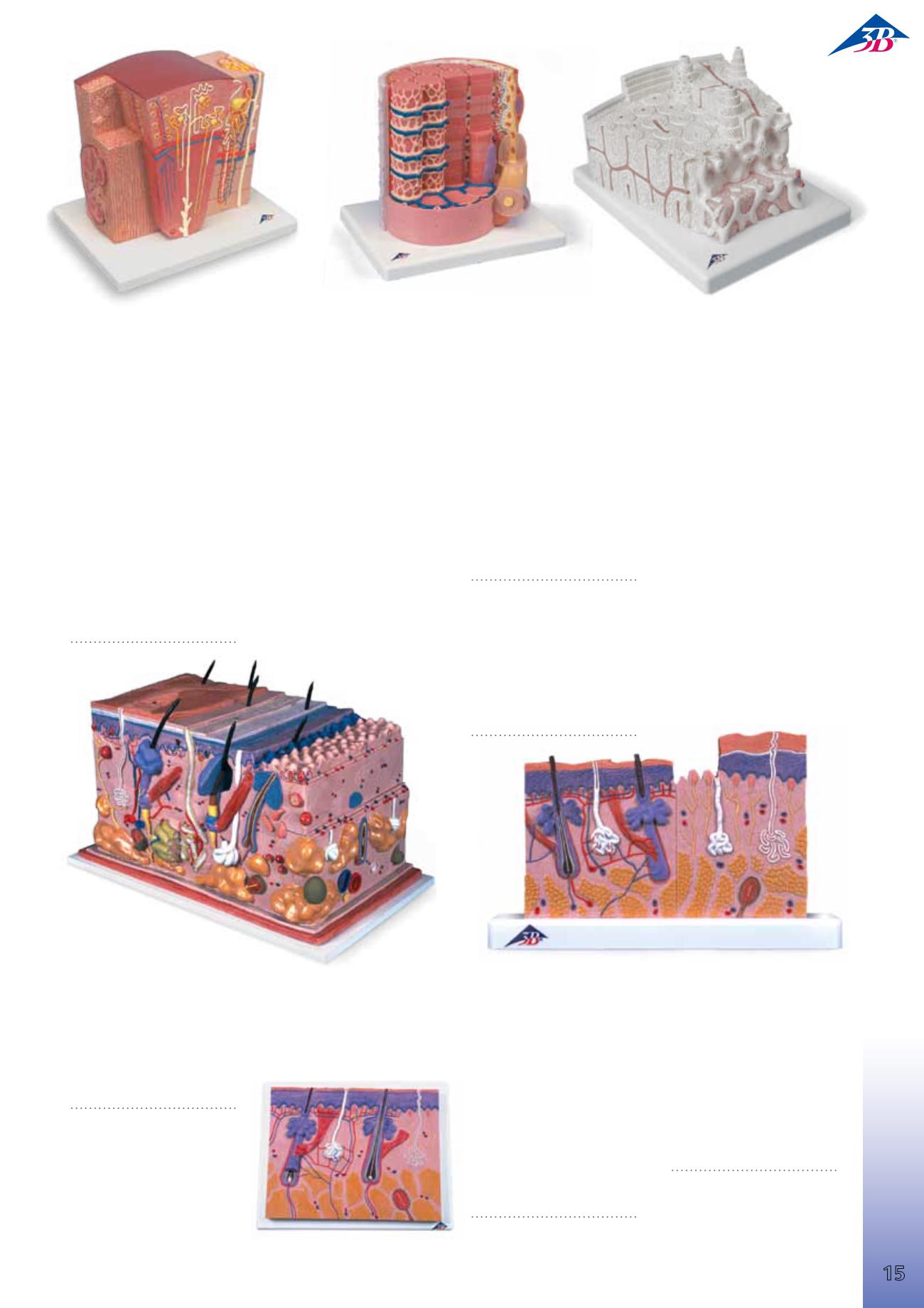
Skin Section, 70 times
full-size
This relief model shows a section
through the three layers of the
hair-covered skin of the head.
Delivered on base it shows:
• Representation of hair follicles
with sebaceous glands
• Sweat glands
• Receptors
• Nerves
• Vessels
26x33x5 cm; 1.0 kg
&
L/E/D/S/F/P/J www.
9985-1000289
Skin Section, 40 times
full-size
The two halves of this relief model
show the three layers of hairy and
hairless skin in order to make the
differences clear. In detail with hair
follicles, sebaceous glands, sweat
glands, receptor, nerves and
vessels. Delivered on base.
24x15x3.5 cm; 0.2 kg
&
L/E/D/S/F/P/J www.
9985-1000290
Skin, Block Model, 70 times full-size
This unique model shows a section of human skin in three dimensional
form. Individual skin layers are differentiated, and important structures
such as hair, sebaceous and sweat glands, receptors, nerves and vessels are
shown in detail. Mounted on baseboard.
44x24x23 cm; 3.6 kg
&
L/E/D/S/F/P/J www.
9985-1000291
3B MICRO
anatomy
™ Bone Structure
This extremely detailed model depicts a three-dimensional section of a la-
mellar bone, showing the typical structure of a tubular bone enlarged 80
times. Various planes are shown in cross and longitudinal section through
all levels of the bone, as well as a 2‑planesection through the inner struc-
ture of the bone marrow. The typical elements of a lamellar bone are easily
identified and help to understand its structure and function with the char-
acteristic osteons, also referred to as Haversian systems. This model allows
a graphic illustration of the interplay of the individual components, such as
spongy and compact substance, endosteum, cortical substance, osteocytes,
Volkmann and Haversian canals.
Supplied on base.
26x19x14.5 cm; 0.8 kg
&
E/D/S/F/P/J www.
9985-1000154
3B MICRO
anatomy
™ Muscle Fibre
The model illustrates a section of a skeletal muscle fibre and its neuro
muscular end plate magnified approx. 10.000 times. The muscle fibre is
the basic element of the diagonally striped skeletal muscle.
23.5x26x18.5 cm; 1.1 kg
&
L/E/D/S/F/P/I/J
9985-1000213


