63
9985-1001261
9985-1005113
9985-1001262
9985-1000230
9985-1005114
9985-1005555
. . . g o i n g o n e s t e p f u r t h e r
Anatomy
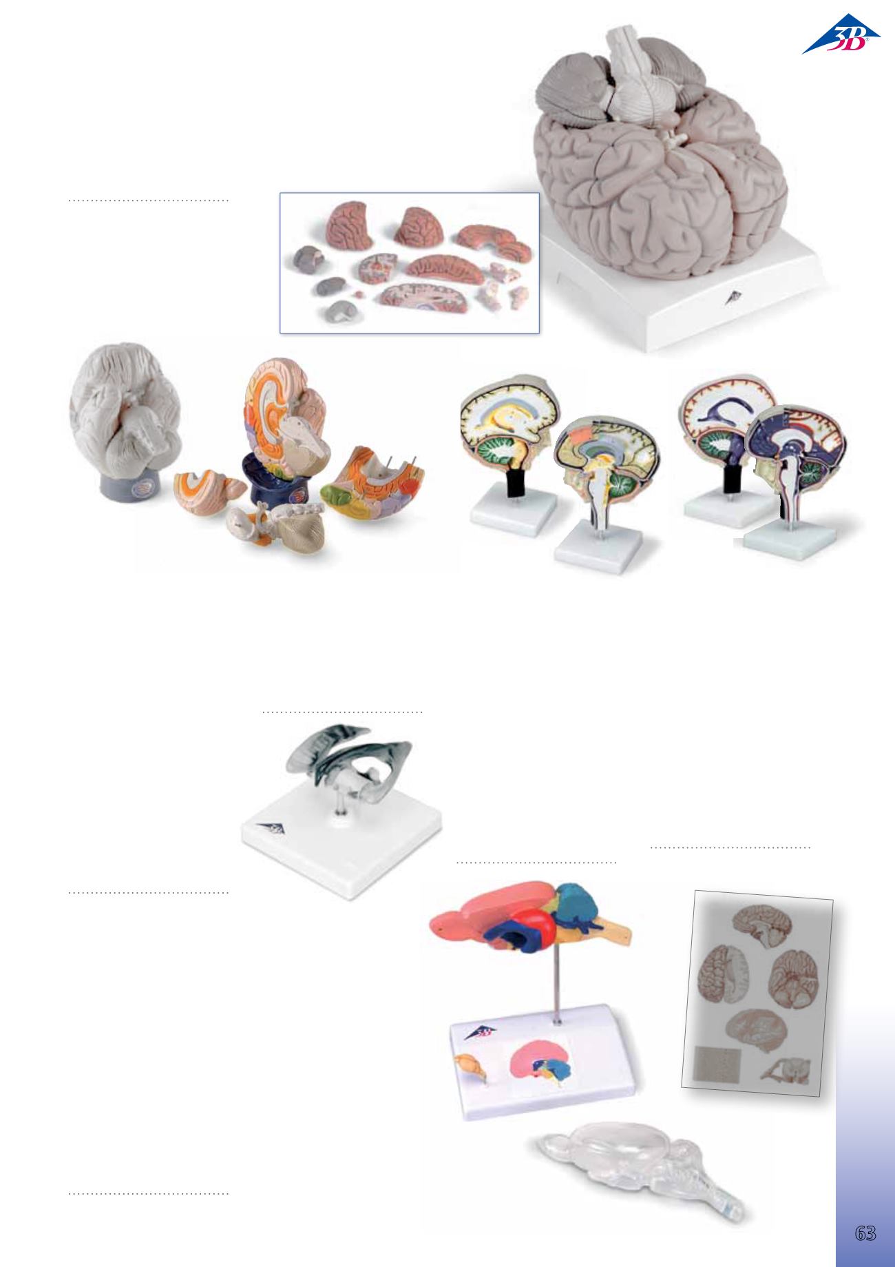
Bra in
Giant Brain, 2.5 times full size, 14-part
A comprehensive brain model that is also a very useful teaching aid, es
pecially for large groups of students. All structures of the brain and the
ventricles are visible through median, frontal and horizontal sections.
Delivered on removable base.
34x30x37 cm; 5.6 kg
L/D/E/F/S
9985-1001261
Cerebrospinal Fluid
Circulation
Enlarged, detailed model of a sec-
tion through the right half of the
brain showing the cut pia mater,
arachnoid and dura mater. The
model has the cerebrospinal fluid
areas clearly identified and the di-
rection of flow indicated by arrows.
Bright colours to distinguish impor-
tant features; identified in English
in an accompanying key card.
Mounted on stand.
25x18x12 cm; 0.9 kg
E
9985-1005114
Brain Section
An enlarged and very detailed sec-
tion through the right half of the
brain, including a portion of the
skull. The pia mater has been re-
moved. This model is double sided
and finely coloured. One surface is
on the median line, including a
section of the falx cerebri. A sagit-
tal cut on the reverse exposes the
lateral ventricle. There are 49 refer-
ences on the model, identified in
English in an accompanying key
card. Mounted on a stand.
25x18x12 cm; 0.9 kg
E
9985-1005113
Regional Brain, 4-part
The following lobes and regions of
this 2-times life-size brain are rep-
resented in different colours and
labeled in English:
• Frontal lobe
• Parietal lobe
• Occipital lobe
• Temporal lobe
• Motor cortex
• Somatosensory cortex
• Limbic cortex
• Cerebellum
• Brain stem
The twelve cranial nerves and addi-
tional features are numbered.
Supplied with wooden stand.
23x20x30 cm; 2.38 kg
E
9985-1005555
Brain Ventricle
This model shows both side ventri-
cles, the 3rd and 4th ventricle and
the Aquaeductus cerebri (Sylvius).
On stand.
14x11x14 cm; 0.6 kg
L/D/E/F/S/P/I/J www.
9985-1001262
Rat Brain Comparative Anatomy
The model shows a rat brain in approx. 6-fold enlargement. Sectioned me-
dially, it can be disassembled into two halves. The right half of the model
shows the structures of the cerebrum, cerebellum and brain stem, each of
which is colour-coded for didactic purposes (cerebrum = pink, cerebellum
= blue, brain stem = yellow), both externally and in the median section.
The left half of the model is largely transparent, thus revealing a view of
the coloured left lateral ventricle and hippocampus, which can also be seen
in the median section. For purposes of comparison, a natural cast of a rat
brain and a didactic, small-scale illustration of a human brain in median
section are shown on the base, with the same colour coding used for the
various regions.
14x10x16 cm; 0.24 kg
L/D/E/F/S/P/I/J www.
9985-1000230
9985-4006536
–
9985-1001185
+


