64
9985-1000238
9985-1000232
9985-1000233
9985-1000234
9985-1000235
9985-1000237
9985-1000236
9985-1005553
3 B S c i e n t i f i c ® M e d i c a l
Anatomy
Ner vous Sys tem
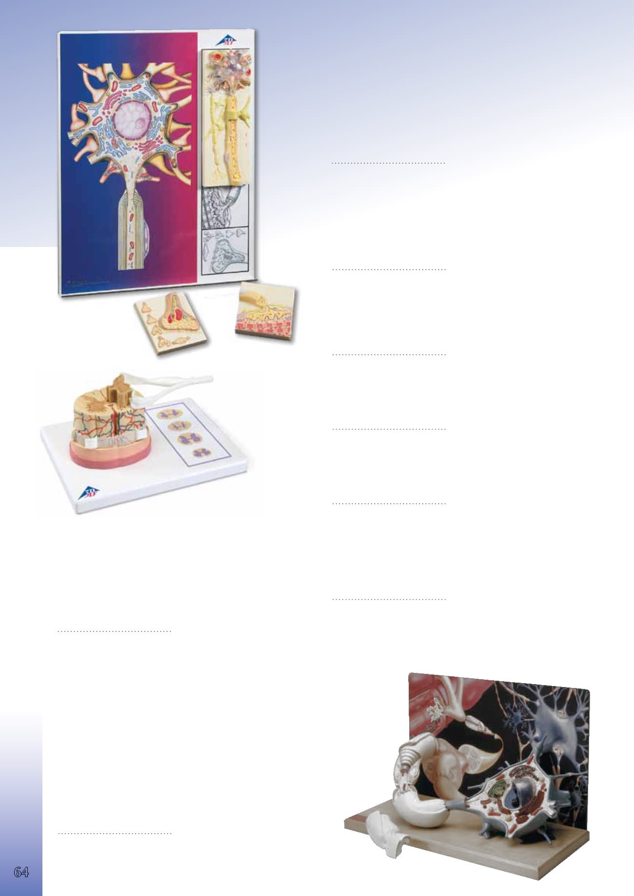
“Physiology of Nerves” Series, 5 Magnetic Models on Illustrated
Metal Board
Displaying the basic structures of the human nervous system. Each of the
five sections shows a plastic coloured relief model of the main synapse
variations. All sections can magnetically attach to the illustrated base which
depicts the neural components in vivid colours. Each section is also availa-
ble separately.
68x51x10 cm; 4.2 kg
E/D/S/F/P www.
9985-1000232
Neuron Cell Body
Typical neuron body with cell organelles, for example mitochondria and
many other characteristics of human cell, are visible through a removable
transparent cover. The edge of the cell body also shows the synapses of
connected neurons.
12.2x11.7x6.2 cm; 0.2 kg
9985-1000233
Myelin Sheaths of the CNS
This model shows the glial cells which build the insulating layer around the
axons of the central nervous system.
12.2x11.7x3.6 cm; 0.2 kg
9985-1000234
Schwann Cells of the PNS
Depicts a Schwann cell with sectioned core.
12.2x11.7x3.2 cm; 0.2 kg
9985-1000235
Motor End Plate
Neuromuscular junction with striated muscle fibre is depicted.
12.0x11.5x3.2 cm; 0.2 kg
9985-1000236
Synapse
Featuring the endoplasmic reticulum, mitochondria and the membranes of
the synaptic gap. Also depicts 5 smaller relief models of the main synapse
variations.
12.0x11.5x2.7 cm; 0.2 kg
9985-1000237
Spinal Cord with Nerve Endings
The model illustrates the composition of the spinal cord, magnified to
a scale of about 5:1. The spinal cord is formed by a central channel sur-
rounded by “grey matter” with an outer layer of “white matter”. The base
features illustrations of various cross sections through the white and grey
matter at the neck, torso, lumbar and sacral regions. Supplied on a base.
26x19x13 cm, 0.4 kg
L/D/E/S/F/P/I/J www.
9985-1000238
Motor Neuron Diorama
Magnified more than 2,500 times, this model represents a fully three
dimensional reproduction of a motor nerve cell situated within a milieu of
interacting neurons and a skeletal muscle fibre. The membranous envelope
has been cut away from the neuron to expose the cytological ultrastructure,
organelles and inclusions within the cell body. Branching dendrites, com-
municating synapses and a myelin wrapped axon with node of Ranvier,
project from the neuronal surface. A section of the axon lifts off to let you
view the tightly wound layers of the enveloping myelin sheath and neuro-
lemma, as well as the Schwann cell which formed them.
Mounted on a wooden base.
43x20x28 cm; 3.0 kg
E
9985-1005553


