40
9985-1005100
9985-1000175
9985-1000003
9985-1002390
9985-1000180
3 B S c i e n t i f i c ® M e d i c a l
Joint s
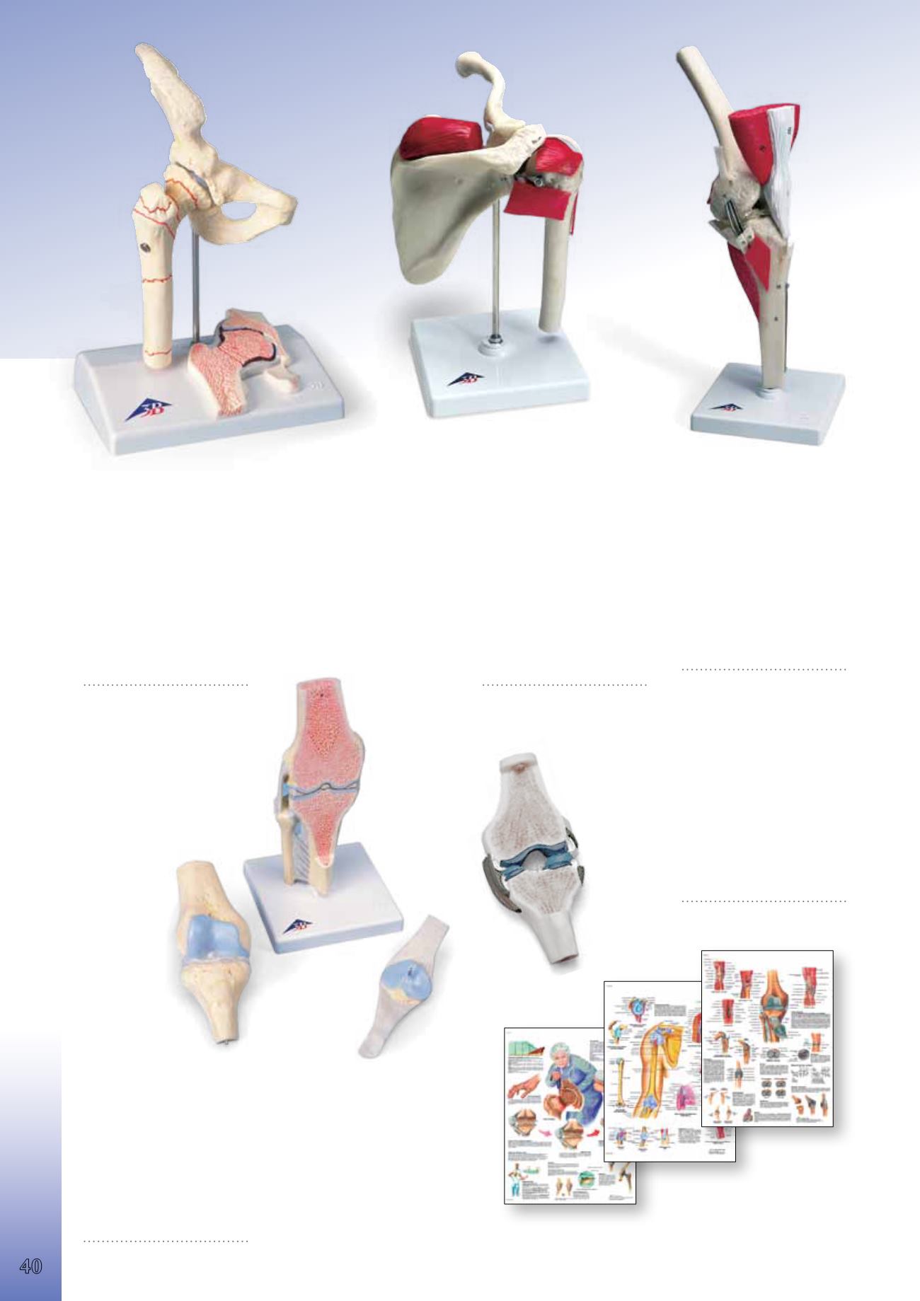
Femoral Fracture and Hip Osteoarthritis
This model was developed to provide patients with understandable infor-
mation, e.g. before surgery. It shows the right hip joint of an elderly person
in half natural size. In addition, a frontal section through the femoral neck
is shown in relief on the base. The model shows the femoral fractures that
occur most commonly as well as typical wear and tear symptoms of the hip
joint. The following fractures are shown: medial femoral neck fracture, lat-
eral femoral neck fracture, fracture through the trochanteric region, frac-
ture below the trochanters, femoral shaft fracture, femoral head fracture,
fracture of the greater trochanter, fracture or avulsion of the lesser
trochanter. Mounted on base. 14x10x22 cm; 0.3 kg
E/D/S/F/P/J www.
9985-1000175
Sectional Knee Joint
Longitudinal section of the human
knee joint. Bone structure, menis-
cus, joint cartilage, synovial mem-
brane and joint ligaments are
shown in colour.
18.5x8.5x5 cm; 0.3 kg
&
E
9985-1005100
Deluxe Knee
Distal half of femur attached to
tibia, fibula and patella. Depicts all
major muscles of the knee. Cruciate/
collateral ligaments simulated with
triple springs. Simulated “Bucket
Handle” tear in medial meniscus.
Patellar tendon simulated.
Stand included.
33x12x12 cm; 0.7 kg
&
E
9985-1002390
Sports Shoulder
Includes upper half of humerus,
clavicle and scapula. Articulated to
show normal movement. Depicts
the following:
• M. supraspinatus
• Long head tendon
• Glenoid labrum
• Rotator cuff
Stand included.
23x17x12 cm; 0.4 kg
&
E
9985-1000003
Sectional Knee Joint Model, 3-part
This model can be used to demonstrate various disorders of the human
knee joint and their respective therapies in a graphic way. The model
shows a natural sized, healthy right knee joint in upright position, includ-
ing parts of the femur, tibia and fibula as well as the ligament system and
the patella with part of the femoral tendon. The patella and attached ten-
don and the front half of the model (which is frontally sectioned) can be
detached. Mounted on base.
12x12x24 cm; 0.5 kg
&
L/E/D/S/F/P/I/J www.
9985-1000180
You will find our large selection
of Charts starting on page 108.
9985-4006654 *
9985-1001474 **
9985-4006658 *
9985-1001482 **
9985-4006661 *
9985-1001488 **
The Knee Joint
Shoulder and Elbow
Arthritis
Anatomy


