38
9985-1000286
9985-1000287
9985-1000288
3 B S c i e n t i f i c ® M e d i c a l
Pe l v i s
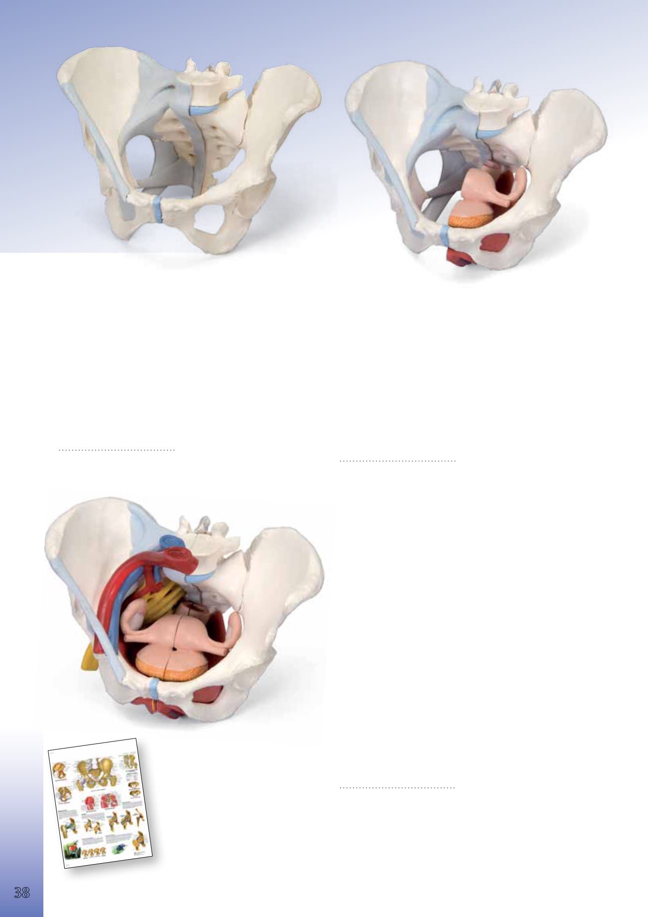
Female Pelvis with Ligaments, 3-part
This three part model represents an original cast of a bony female pelvis,
which shows all details of anatomical structures. Two hip bones, the pubic
symphysis, the sacrum and the coccyx, the fifth lumbar vertebra with in-
tervertebral disc. A midsagital section through the fifth lumbar vertebra,
sacrum and coccyx, allow both halves of the pelvis to be disassembled re-
vealing a part of the cauda equina in the vertebral canal. The left half of
the fifth lumbar vertebra is removable. The right half of the model shows
following pelvic ligaments: inguinal ligament, sacrotuberous ligament,
sacrospinous ligament, anterior sacroiliac ligaments, iliolumbar ligament,
anterior longitudinal ligament, interosseous sacroiliac ligament, posterior
sacroiliac ligament and obturator membrane.
19x27x19 cm, 1,0 kg
&
L/D/E/F/I/S/P/J/R/C www.
9985-1000286
Female Pelvis with Ligaments,
Vessels, Nerves, Pelvic Floor and Organs, 6-part
This six part model of a female pelvis represents detailed information about
the topography of bones, ligaments, vessels, nerves, pelvic floor muscles
and female pelvic organs. It presents the whole pelvic floor with partially
removable midsagitally sectioned external anal sphincter, external urethral
sphincter, deep and superficial transverse perineal and bulbospongiosus.
Rectum, uterus with fallopian tubes ovaries and vagina are also removable
and can be disassembled into 2 halves by midsagital section. The right pel-
vic half demonstrates the divisions and topographical anatomy of the com-
mon iliac artery, the external and internal artery and also of the common
iliac vein and the external iliac vein. The right sacral plexus, right sciatic
nerve and right pudendal nerve are also shown. Bones and ligaments pre-
sented: Two hip bones, the pubic symphysis, the sacrum and the coccyx, the
fifth lumbar vertebra with intervertebral disc. A midsagital section through
the fifth lumbar vertebra, sacrum and coccyx, allow both halves of the pel-
vis to be disassembled revealing a part of the cauda equina in the vertebral
canal. The left half of the fifth lumbar vertebral body is removable. The
right half of the model shows following pelvic ligaments: inguinal ligament,
sacrotuberous ligament, sacrospinous ligament, anterior sacroiliac liga-
ments, iliolumbar ligament, anterior longitudinal ligament, interosseous
sacroiliac ligament, posterior sacroiliac ligament and obturator.
19x27x19 cm, 1,6 kg
&
L/D/E/F/I/S/P/J/R/C www.
9985-1000288
Female Pelvis with Ligaments, Midsagitally Sectioned
through Pelvic Floor Muscles Organs, 4-part
This four part model of a female pelvis represents detailed information
about the topography of bones, ligaments, pelvic floor muscles and female
pelvic organs. The right half shows the bones with pelvic ligaments.
In addition, the left half of the pelvis contains the muscles of the pelvic
floor including levator ani, ischiocavernosus, deep and superficial trans-
verse perineal, external anal sphincter, external urethral sphincter. A par-
tially removable bulbospongiosus demonstrates the vestibular bulb and
Bartholin gland. The removable midsagital section through the urinary
bladder, vagina, uterus and rectum demonstrates the relationship of the
muscles of the pelvic floor to the openings for urethra, vagina and rectum.
19x27x19 cm, 1,3 kg
&
L/D/E/F/I/S/P/J/R/C www.
9985-1000287
You will find our large selection
of Charts starting on page 108.
9985-4006660 *
9985-1001486 **
Pelvis and Hip
Anatomy


