36
9985-1000182
9985-1005326
9985-1005866
3 B S c i e n t i f i c ® M e d i c a l
Spina l di sorder s
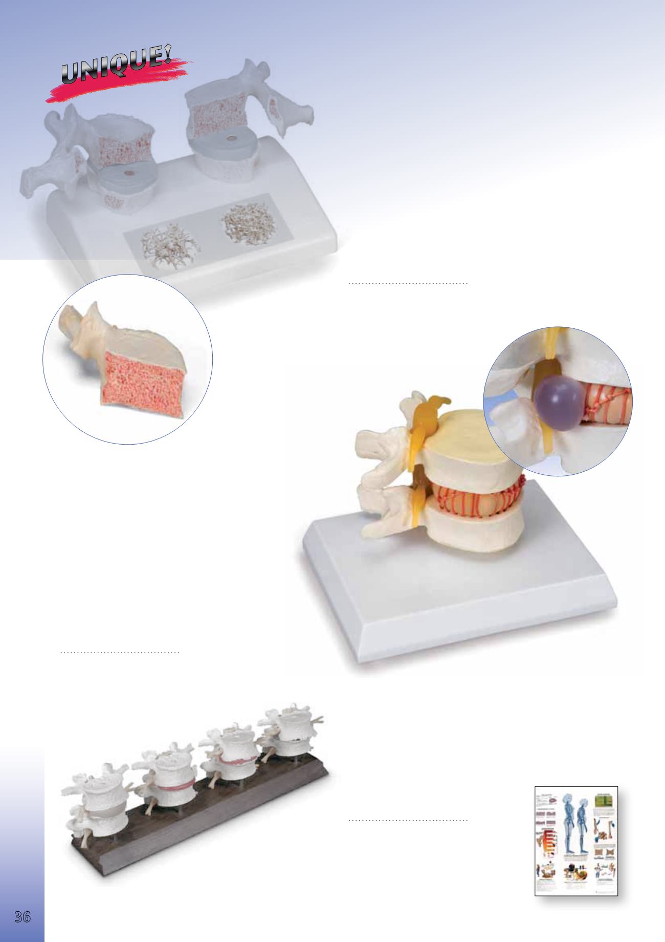
Herniated Disc Simulator
This innovative model shows the injured mechanism of a
herniated disc. The simulator demonstrates how the in-
tervertebral disc prolapses when flexing the vertebrae, re-
producing what happens in real life when we bend or twist
our trunk. Since vertebral disorders are widespread, thera-
pists should have this model to make people aware of the
importance of gentle movements and behaviours for the
spinal column. This model will be of great assistance in
medicine, physiotherapy, medical surgeries and clinics,
moving and handling, ergonomics, physical education and
other fields. The model consists of two vertebrae with an
elastic intervertabral disc, spinal cord and nerves.
Size: 12x11.5x9 cm; weight 0.6 kg
9985-1005326
Osteoporosis Model
Impressive didactic model for comparing osteoporotic and normal thoracic
vertebrae. Ideal for medical studies and patient education.The 11th and
12th thoracic vertebrae are shown. Reproductions of sequential osteo
porotic thoracic vertebrae with narrower intervertebral disc are located on
the left of the stand. The upper vertebra is divided in the middle.The mag-
netically attached vertebral half can be removed easily to show the cut sur-
faces.This allows clear visualisation of the fractured upper part of the verte-
bral body caused by sintering, i.e. collapse of the bony substance in the
course and as a result of osteoporosis.Degenerative changes in the bone,
manifested as osteophytes, are also identifiable. For comparison, reproduc
tions of two corresponding healthy vertebrae with intervertebral disc are
provided on the right side. One half of the upper vertebral body is also
magnetically attached and can be removed. Each set of vertebrae also has
a sticker on the stand showing two 3D micro CT images obtained from bone
biopsies. These illustrate the microarchitecture of the osteoporotic bone,
which has a lower bone density compared to healthy bone.
14x9x10 cm; 0.2 kg
&
L/D/E/F/I/S/P/J/R/C www.
9985-1000182
4-Stage Degenerative Lumbar Set
An exceptional model demonstrating bone and disc degeneration. The
vertebrae pairs (L4, L5) demonstrate from left to right: a normal disc and
bone; Facet Syndrome and a herniated disc; thinning disc and the begin-
ning of bone spurring; a seriously degenerated disc with bone fusing.
Mounted on base.
8.5 cm, 0.5 kg
&
E
9985-1005866
You will find our large
selection of Charts
starting on page 108.
Osteoporosis
9985-4006653 *
9985-1001472 **
Anatomy


